Jul 01,2022
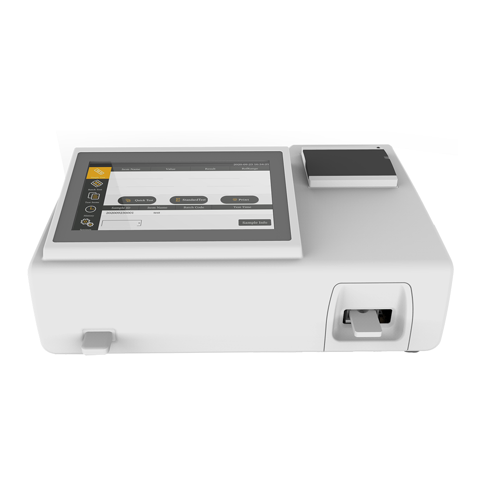
Fluorescence immunoassay analyzers employ several strategies to minimize background noise and improve signal detection, particularly in complex samples where non-specific interactions and autofluorescence can obscure the target signal. Here’s how these analyzers achieve improved accuracy and sensitivity:
1. Optimized Wavelength Selection:
Emission and Excitation Filters: Fluorescence analyzers are equipped with highly specific optical filters that select the precise wavelengths for both excitation and emission. This reduces the chances of background noise from other fluorescent materials or light sources. By using narrow bandpass filters, the system can selectively excite the fluorophore and detect only the emitted light at the target wavelength, minimizing interference.
Multiple Channels: Many fluorescence analyzers use multiple detection channels, each optimized for different fluorophores. This allows for simultaneous detection of multiple targets in a sample (multiplexing) while minimizing cross-talk between signals.
2. Fluorophore Choice and Brightness:
High-Performance Fluorophores: Selecting fluorophores with high quantum yield (efficiency of photon emission) and narrow emission spectra is key to reducing background noise. Bright fluorophores provide stronger signals, making them less susceptible to interference from background fluorescence in the sample.
Stability of Fluorophores: Stable fluorophores that are less prone to photobleaching (degradation under light exposure) help maintain consistent signal strength and reduce false negatives that might arise due to weakened signals over time.
3. Time-Resolved Fluorescence (TRF):
Time-Resolved Detection: Some fluorescence immunoassay analyzers incorporate time-resolved fluorescence (TRF), a technique where the emission signal is detected after a brief delay following the excitation pulse. This allows for the detection of signals from fluorophores that exhibit delayed emission (like lanthanide chelates) while avoiding interference from background fluorescence that occurs immediately after excitation.
Time-Gated Detection: This technique helps distinguish the true signal from background noise by gating the detector to collect light only during the time period when the target fluorophore emits its characteristic signal. Background noise typically decays rapidly, whereas the specific signal from the target fluorophore remains detectable over a longer time frame.
4. Background Subtraction and Signal Processing:
Advanced Software Algorithms: Modern fluorescence analyzers employ sophisticated algorithms to subtract background fluorescence from the final readout. These algorithms analyze the raw signal, identifying the background fluorescence levels, and subtract them from the measured signal to yield a more accurate result.
Dynamic Range Compression: Signal processing can also involve compressing the dynamic range of the detector, ensuring that low and high signals are both accurately measured. This is especially useful in complex samples where the difference between background and target signal is minimal.

5. Optical Path Design and Shielding:
Optical Path Isolation: Fluorescence analyzers often feature carefully designed optical paths that isolate the detector from stray light and scatter. Optical components, such as beam splitters, mirrors, and lenses, are precisely aligned to ensure that only light emitted by the fluorophores reaches the detector, minimizing ambient light interference.
Light Shielding and Enclosures: The sample chamber is often enclosed or shielded to prevent ambient light or room fluorescence from affecting the readings. This ensures that the detector receives light only from the fluorescence signals generated by the assay.
6. Sample and Reagent Control:
Proper Sample Preparation: Ensuring that samples are properly prepared and free of contaminants that could fluoresce is crucial. This may involve optimizing the concentration of analytes, reducing matrix effects (interference from sample components), and removing background substances that could emit unwanted fluorescence.
Blocking and Quenching: Blocking agents or quenching reagents are sometimes used to minimize non-specific binding and reduce background fluorescence from unbound antibodies or other components in the sample. For example, quenching can reduce autofluorescence from certain sample types (e.g., biological tissues or blood).
7. Auto-calibration and Calibration Standards:
Automated Calibration: Many analyzers come with automated calibration systems that help maintain optimal signal-to-noise ratios over time. The system can adjust parameters such as gain, exposure time, and detection thresholds based on calibration standards that minimize the impact of background noise.
Standardization with Controls: Regular use of internal and external control samples allows for the detection of any drift or changes in system performance that might contribute to increased background noise, ensuring consistent, accurate results.
8. Use of High-Quality Optics and Detection Systems:
Advanced Detectors (e.g., PMTs or CCDs): Fluorescence analyzers often utilize photomultiplier tubes (PMTs) or charge-coupled devices (CCDs) with high sensitivity and low noise characteristics to detect weak signals. These detectors are engineered to maximize signal detection while minimizing background interference.
Amplification and Filtering: Advanced detectors are often paired with signal amplification and low-pass filtering systems that ensure the true fluorescence signal is detected and amplified without amplifying noise.
9. Minimizing Autofluorescence from Sample Matrices:
Sample Pre-Treatment: Some analyzers use specific protocols to reduce autofluorescence in complex sample matrices, such as biological tissues or serum. These pre-treatment steps might involve the use of specific buffers or chemical agents to reduce the inherent fluorescence of the sample, allowing for a cleaner signal from the target fluorophore.


 Español
 Français
 Deutsch
 عربى
عربى


